

Dental X-rays play an important role in any dental treatment plan. There are a variety of different examinations, preventative and diagnostic, that allow clinicians a better understanding of each patient’s anatomy and needs. Dental imaging helps clinicians diagnose common problems such as gum disease, tooth and gum infections, cavities, and gauge bone density. Without imaging to supplement dental consultations, some conditions may be left undiagnosed and, subsequently, untreated.
Dental imaging falls into two categories: intraoral and extraoral.
Intraoral
Intraoral examinations are captured inside your mouth. They help clinicians visualise cavities, tooth roots and developing teeth at specific sites with very high detail. Intraoral examinations often include the following imaging:
- Bitewing X-rays – to check in-between teeth, or assess restorations
- Periapical X-rays – to gain close-up views of individual teeth, including their roots
- Occlusal – imaging to assess the bite relationship between upper and lower teeth
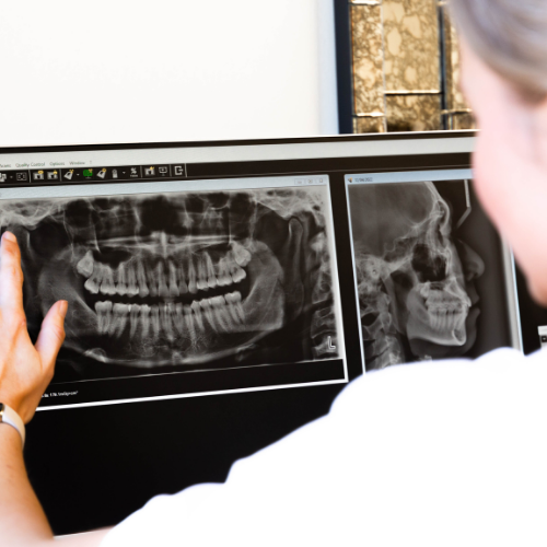
Extraoral
Extraoral examinations are captured outside of your mouth. They cover a larger area and provide great detail of all of your teeth, jaw and oral anatomy in one exposure. Extraoral examinations often include the following imaging:
- Orthopantomogram (OPG) – a full mouth X-ray
- Cephalometric imaging – X-rays of the head and facial profile
- Cone Beam Computed Tomography (CBCT) – 3D X-ray imaging of necessary areas of the face and teeth
Great care is taken to ensure dental X-rays are taken only when necessary and are kept to a minimum. This helps keep the radiation dose low should you receive multiple x-rays during your dental treatment.
If your dentist requires you to have an X-ray, they will select the most appropriate type of imaging for your needs. If an extraoral radiograph is required, you may be referred to an imaging clinic outside of the dental practice. In these cases, a radiographer will perform your imaging ensuring the exam uses the lowest dose possible while still providing a high-quality image. Radiation safety is taken very seriously with any X-ray examination, including exposure selection, collimation (the precision of the radiation beam) and lead shielding.
We are exposed to radiation everyday in our own natural environment. The amount of natural background radiation a person receives is variable and dependent on environmental factors like soil, rock, altitude and latitude in conjunction with your diet. The average amount of background radiation exposure in Australia for any one person currently sits at approximately 1.5 mSv per year from natural sources, a near equivalent to 75 chest X-rays. Conventional 2D dental X-rays emit very small doses of radiation and are deemed to be very safe.
Digital X-rays use very low doses of radiation, with dental imaging using the lowest dose of all plain-film radiography. A person would receive between 0.001-0.005mSv from a single dental exposure, emitting the same amount of radiation you would typically receive on a one- to two-hour flight, or comparable with one day of natural background radiation exposure. Radiation becomes harmful at very high doses (over 500mSV) and reactions occur very shortly after exposure. These high doses are only experienced as a result of large nuclear or radiation accidents, definitely not from plain film radiography.
If you have any concerns regarding your dental imaging, your dentist or radiographer will be more than happy to discuss this with you prior to your imaging.


