

Most jaw pain treatment fails because the diagnosis was based on guesswork. Here is what data-driven TMJ diagnosis technology looks like – and why it changes outcomes.
There is a reason that so many patients with jaw problems feel like they have been going around in circles. They see a dentist who says they grind their teeth and makes a night guard. They see a physiotherapist who treats their neck. They see their GP who prescribes anti-inflammatories. Each practitioner addresses one piece of the puzzle based on their individual examination. None of them has the full picture.
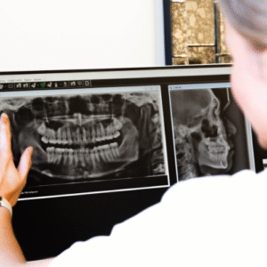
The fundamental challenge with diagnosing temporomandibular disorders (TMD) is that clinical examination alone – even by an experienced practitioner – has significant limitations. You can palpate muscles for tenderness, but you cannot feel which ones are overactive and by how much. You can check the bite with articulating paper, but you cannot see the timing and magnitude of forces across every tooth simultaneously. You can take a standard X-ray, but you cannot visualise the three-dimensional anatomy of the joint, the airway, or the disc.
At Smile Solutions, our TMD Clinic has invested in TMJ diagnosis technology that moves beyond clinical judgement into objective, measurable data. EMG muscle mapping, T-Scan bite force analysis, CBCT imaging, and thermal assessment provide a comprehensive diagnostic picture that guides treatment with a precision that was simply not possible a generation ago. This article takes you inside that diagnostic process.
EMG Muscle Mapping: Seeing What Muscles Are Doing
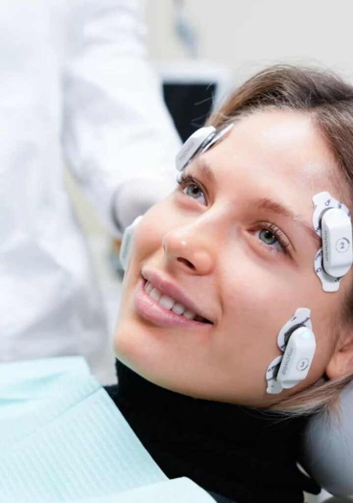
Electromyography (EMG) measures the electrical activity generated by muscles when they contract. In our TMD Clinic, we use surface EMG – non-invasive electrodes placed on the skin over the muscles of mastication – to record and analyse muscle function in real time.
What we measure:
Resting muscle activity
In a healthy jaw, the muscles of mastication should be relatively quiet at rest – active enough to maintain jaw position but not generating significant contractile force. In many TMD patients, one or more muscles show elevated resting activity, indicating chronic tension or guarding. The EMG quantifies exactly which muscles are overactive and by how much.
Functional muscle activity
During clenching, chewing, and jaw movements, the muscles should fire in coordinated, symmetrical patterns. EMG reveals whether the left and right masseter muscles are balanced, whether the temporalis and masseter activate in the correct sequence, and whether any muscle is working disproportionately hard to compensate for dysfunction elsewhere.
Muscle symmetry
Left-right asymmetry in muscle activity is a hallmark of many TMD conditions. A patient clenching predominantly on the right side will show significantly higher masseter EMG readings on the right. This asymmetry may correlate with a bite discrepancy, joint problem, or postural deviation that needs to be addressed.
Fatigue patterns
By recording muscle activity over sustained clenching, we can assess how quickly muscles fatigue and whether fatigue patterns differ between sides. Abnormal fatigue patterns may indicate chronic overload, metabolic compromise within the muscle, or compensatory loading.
How this changes treatment:
EMG data transforms splint design from an educated guess into a targeted intervention. If the right masseter is hyperactive at rest while the left temporalis is showing compensatory overactivity, the splint can be designed and adjusted to specifically address those imbalances. Without EMG, the splint design is based on anatomical assumptions rather than functional data.
EMG also provides an objective baseline against which treatment progress can be measured. After wearing a splint for six weeks, we can repeat the EMG assessment and quantify whether muscle activity has normalised. This is fundamentally different from asking the patient “Does it feel better?” – which, while important, is subjective and can be influenced by many factors.
T-Scan Bite Force Analysis: The Dynamic Occlusal Map
The T-Scan system is a digital occlusal analysis tool that records the location, timing, and magnitude of tooth contacts during biting and chewing. A paper-thin sensor is placed between the teeth, and as the patient closes into maximum intercuspation (full bite), the system generates a real-time, three-dimensional force map.
What T-Scan reveals that articulating paper cannot:
Force magnitude at each contact point
T-Scan does not just show where teeth touch – it shows how much force each contact carries. A contact that appears small on articulating paper might be bearing 40 per cent of the total bite force. This disproportionate loading can drive muscle compensation, joint overloading, and tooth fracture.
Timing sequence
When you close your teeth together, ideally all teeth should contact simultaneously (or within a few milliseconds of each other). T-Scan reveals the sequence of contacts with millisecond precision. A tooth that contacts 50 milliseconds before the others (a premature contact) acts as a fulcrum, triggering reflexive muscle responses and potentially driving clenching behaviour.
Centre of force trajectory
As the teeth come together, the centre of force should move smoothly to a central, balanced position. T-Scan displays this trajectory graphically, revealing asymmetries and instabilities in the occlusion that may not be detectable by clinical examination.
Disclusion time
During lateral jaw movements (moving the jaw to the side), the teeth on the non-working side should disocclude (separate) quickly. Prolonged contact on the non-working side during lateral excursion increases lateral pterygoid muscle activity and can contribute to TMD symptoms. T-Scan measures disclusion time precisely, guiding occlusal adjustment to reduce this mechanical trigger.
How this changes treatment:
T-Scan data allows us to perform occlusal adjustments with surgical precision. Rather than empirically grinding down high spots based on articulating paper marks, we can identify the specific contacts that are carrying excessive force or creating timing imbalances and adjust only those points. The result is minimal, targeted tooth modification that produces maximal functional improvement.
For splint therapy, T-Scan data guides the adjustment of the splint’s occlusal surface. We can verify that the splint provides even, simultaneous contacts across all teeth and eliminate premature contacts or force concentrations that might perpetuate muscle dysfunction.
CBCT Imaging: The Three Dimensional View
Cone beam computed tomography (CBCT) provides three-dimensional radiographic imaging at a fraction of the radiation dose of conventional medical CT scanning. In our TMD Clinic, CBCT serves multiple diagnostic purposes.
TMJ bony anatomy
CBCT allows detailed assessment of the condylar (jaw joint) morphology, articular eminence shape, joint space dimensions, and any degenerative changes (osteophytes, flattening, erosion, sclerosis). This information is critical for distinguishing between muscular TMD (which may have normal-appearing joints) and arthrogenic TMD (where the joint itself is the problem).
Condylar position
By measuring the joint space on both sides, we can assess whether the condyles are seated symmetrically or whether one is displaced anteriorly, posteriorly, or laterally. Condylar displacement correlates with disc position and can guide splint design.
Airway assessment
This may be the most important application of CBCT in the TMD Clinic context. The relationship between sleep-disordered breathing and TMD – particularly bruxism – is now well-established. CBCT airway imaging allows us to:
- Measure the minimum cross-sectional area of the upper airway
- Identify the level and nature of any airway narrowing
- Assess the tongue position relative to the airway
- Evaluate the soft palate length and thickness
- Identify anatomical risk factors for obstructive sleep apnoea
This airway data is critical because it helps us determine whether a patient’s bruxism might be driven by airway compromise rather than stress or occlusal factors. If the airway is significantly narrow, a mandibular advancement splint (designed to hold the jaw forward and open the airway) may be more appropriate than a standard stabilisation splint.
Dental and skeletal assessment
CBCT also provides detailed imaging of the teeth, roots, and surrounding bone, allowing assessment of periodontal status, root pathology, and skeletal relationships that may be relevant to the TMD presentation.
Thermal Imaging: Detecting Inflammation
Infrared thermal imaging captures the temperature distribution across the face, highlighting areas of increased blood flow and inflammation. In the context of TMD assessment, thermal imaging can:
- Identify areas of acute inflammation around the TMJ
- Detect asymmetries in blood flow between the left and right sides
- Visualise vascular patterns associated with muscle hyperactivity
- Monitor changes in inflammatory patterns over the course of treatment
While thermal imaging is not diagnostic in isolation, it adds another objective data point to the comprehensive picture. An area of increased temperature over a tender TMJ confirms that inflammatory processes are active, guiding decisions about anti-inflammatory management and activity modification.
Putting It All Together: The Integrated Diagnostic Picture
The power of our diagnostic approach lies not in any single technology but in the integration of multiple data streams into a coherent clinical picture.
Consider a patient who presents with right-sided jaw pain and morning headaches. Traditional examination finds tenderness in the right masseter and clicking in the right TMJ. A standard night guard might be prescribed based on these findings.
Our comprehensive assessment reveals:
- EMG: Right masseter resting activity is three times higher than left; right temporalis shows compensatory hyperactivity
- T-Scan: A premature contact on the upper right second molar is carrying 35 per cent of total bite force and contacts 40 milliseconds before any other tooth
- CBCT: The right condyle shows early degenerative changes with reduced joint space; the airway cross-section is narrowed at the level of the tongue base
- Thermal imaging: Elevated temperature over the right TMJ consistent with active inflammation
This integrated picture tells a completely different story from the simple examination findings. The premature contact is driving asymmetric muscle loading, which is overloading the right TMJ and contributing to degeneration. The airway narrowing suggests possible sleep-disordered breathing contributing to nocturnal clenching. The treatment plan can now address each factor specifically:
- Occlusal adjustment to eliminate the premature contact
- A splint designed to reduce right-sided loading based on EMG and T-Scan data
- Referral for sleep study based on airway findings
- Anti-inflammatory management based on thermal and CBCT joint findings
- Osteopathic treatment with our in-house osteopath Rachel Smith for muscular management
This is the difference between treating a symptom and treating a condition. The technology does not replace clinical judgement – it arms clinical judgement with data that makes better decisions possible.
The Outcome Difference
Patients who receive data-driven TMD diagnosis and treatment consistently report better outcomes than those treated empirically. This is not surprising. When you know exactly what is wrong – which muscles are overactive, which tooth contacts are problematic, whether the joint is degenerating, whether the airway is compromised – you can treat with precision rather than probability. We also use these technologies to monitor treatment progress objectively. Rather than relying solely on patient-reported symptom improvement (which is important but subjective), we can re-measure EMG activity, re-scan force distribution, and re-image the airway to verify that treatment is producing measurable physiological changes.
Your Next Steps
If you have been struggling with jaw pain, headaches, clicking, or grinding, and previous treatments have not provided lasting relief, the diagnostic approach may be the missing piece.
Our TMD Clinic at Smile Solutions offers the comprehensive, technology-supported assessment described in this article.